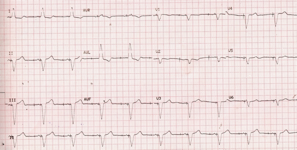
First Degree Pacemaker Exit Block
Abstract
Usually atrial and ventricular depolarizations follow soon after the pacemaker stimulus (spike on the ECG). But there can be an exit block due to fibrosis at the electrode - tissue interface at the lead tip. This can increase the delay between the spike and atrial or ventricular depolarization. The ECG (Figure 1) illustrates a first degree pacemaker exit block in the atrial lead. The interval between the atrial spike and the P wave is about 120 ms. The interval between the ventricular spike and the QRS complex is just above 40 ms.References
1. Kaul R, Kaul UA, Gupta MP. Wenckebach and Mobitz type II pacemaker exit block. A case report. Indian Heart J. 1980;32:60-2.
2. Sathyamurthy I, Krishnaswami S, Sukumar IP. Hyperkalaemia induced pacemaker exit block. Indian Heart J. 1984;36:176-7.
3. Bashour TT. Spectrum of ventricular pacemaker exit block owing to hyperkalemia. Am J Cardiol. 1986;57:337-8.
4. Varriale P, Manolis A. Pacemaker Wenckebach secondary to variable latency: an unusual form of hyperkalemic pacemaker exit block. Am Heart J. 1987;114:189-92.

Published
2016-10-01
How to Cite
FRANCIS, Johnson.
First Degree Pacemaker Exit Block.
BMH Medical Journal - ISSN 2348–392X, [S.l.], v. 3, n. 4, p. 113-114, oct. 2016.
ISSN 2348-392X.
Available at: <https://www.babymhospital.org/BMH_MJ/index.php/BMHMJ/article/view/100>. Date accessed: 25 apr. 2024.
Section
Images

